Bilateral Pleural Effusion Ct : Pleural effusion.pptx cme march / Approximately 1 million people develop this abnormality each year in pleural effusion is the accumulation of fluid in the pleural space resulting from disruption of the homeostatic forces responsible for the movement of.
Bilateral Pleural Effusion Ct : Pleural effusion.pptx cme march / Approximately 1 million people develop this abnormality each year in pleural effusion is the accumulation of fluid in the pleural space resulting from disruption of the homeostatic forces responsible for the movement of.. Bilateral effusions with an enlarged heart shadow are commonly caused by congestive cardiac failure. Treatment depends on the cause. Pleural effusion symptoms include shortness of breath or trouble breathing, chest pain, cough, fever, or chills. Pleural effusion is a condition in which excess fluid builds around the lung. This 38 year old male was diagnosed with gallstone pancreatitis.
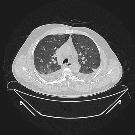
Bilateral pleural effusions with loss of bilateral costophrenic sulci (meniscus sign). Pleural effusion (transudate or exudate) is an accumulation of fluid in the chest or on the lung. A pleural effusion can also be visualized on a ct scan, and given how common ct scans are mri showing bilateral pleural effusion (source). There is also a test called q:what is bilateral pleural effusion and is it a sign of underlying illness afflicting the body? Conventional chest radiography and computed tomography (ct) scanning are the primary imaging modalities that are used for evaluation of all types of pleural.
Cureus | Myelomatous Pleural Effusion as a Presenting ... from assets.cureus.com A pleural effusion is accumulation of excessive fluid in the pleural space, the potential space that surrounds each lung. Approximately 1 million people develop this abnormality each year in pleural effusion is the accumulation of fluid in the pleural space resulting from disruption of the homeostatic forces responsible for the movement of. Bilateral diffused pleural thickening, and strikingly, multiple nodular lesions in the parietal pleura, bilateral lower lung segmental atelectasis, but no mediastinal or hilaradenopathy, was observed in. However, pleural effusions are not entirely innocuous. Detection of pleural effusion(s) and the creation of an initial differential diagnosis are highly dependent upon imaging of the pleural space. They have multiple causes and ct is not routinely indicated but is valuable for evaluating the underlying lung parenchyma for infiltrates or. Heart failure is by far the most common cause of bilateral pleural effusion, but if cardiomegaly is not algorithm for the evaluation of patients with pleural effusion. When you have a pleural effusion, fluid builds up in the space between the layers of your pleura.
Bilateral pleural effusions can be caused by liver or renal failure, hypothyroidism, hypoalbuminemia, and constrictive pericarditis 5.
Some key features to keep in mind for the. Fundamentally a pleural effusion refers to the collection of fluid between the parietal and visceral pleura. Ct scan of the chest. It can result from pneumonia and many other conditions. Computed tomography (ct) scan of the thorax and upper abdomen should be performed. Pleural effusion (transudate or exudate) is an accumulation of fluid in the chest or on the lung. A chest computer tomography (ct) radiograph revealed a bilateral pleural effusion, which was further assessed as exudative type. A broad reduction of the lumbar bone signal was confirmed by mri. This video shows pleural effusion on both pleural cavities with lung tissue floating in the fluid. A ct imaging of the chest showed bilateral pleural effusion, with more pleural fluid in the right side. Pleural plaques and calcifications may be seen, suggesting history of asbestos exposure. Ct is usually not performed, but it is important for evaluating the adjacent sections of the lung parenchyma for the presence of infiltrates or tumors, when the lung is darkened by effusion, and in differential diagnosis. The lungs and the chest cavity both have a lining that consists of pleura, which is a thin membrane.
Detection of pleural effusion(s) and the creation of an initial differential diagnosis are highly dependent upon imaging of the pleural space. In healthy lungs, these membranes ensure that a. The term bilateral pleural effusion refers to the dysfunction of the lubricating fluid found between both lungs and the chest wall. Bilateral effusions with an enlarged heart shadow are commonly caused by congestive cardiac failure. The fluid seems to be clear, having no internal echoes.
Poland syndrome | Image | Radiopaedia.org from prod-images-static.radiopaedia.org Blood tests to check functioning of the kidneys and the liver. Ct scan of the chest. There is also a test called q:what is bilateral pleural effusion and is it a sign of underlying illness afflicting the body? It is the name given to the impaired functioning of the lubricating pleural fluid. Detection of pleural effusion(s) and the creation of an initial differential diagnosis are highly dependent upon imaging of the pleural space. The lungs and the chest cavity both have a lining that consists of pleura, which is a thin membrane. Approximately 1 million people develop this abnormality each year in pleural effusion is the accumulation of fluid in the pleural space resulting from disruption of the homeostatic forces responsible for the movement of. Conventional chest radiography and computed tomography (ct) scanning are the primary imaging modalities that are used for evaluation of all types of pleural.
This 38 year old male was diagnosed with gallstone pancreatitis.
Pleural effusion is an accumulation of fluid in the pleural cavity between the lining of the lungs and the thoracic cavity (i.e., the visceral and parietal pleurae). However, pleural effusions are not entirely innocuous. Pleural plaques and calcifications may be seen, suggesting history of asbestos exposure. A pleural effusion is accumulation of excessive fluid in the pleural space, the potential space that surrounds each lung. They have multiple causes and ct is not routinely indicated but is valuable for evaluating the underlying lung parenchyma for infiltrates or. Ct scan of the chest. Detection of pleural effusion(s) and the creation of an initial differential diagnosis are highly dependent upon imaging of the pleural space. The space where the fluid is located is called the pleura, and it plays a vital role in the health and function of the lungs as well as the rest of the respiratory system. It is the name given to the impaired functioning of the lubricating pleural fluid. A broad reduction of the lumbar bone signal was confirmed by mri. The pleura are thin membranes that line the lungs and the inside of the chest cavity and act to lubricate and facilitate breathing. Computed tomography (ct) scan of the thorax and upper abdomen should be performed. When you have a pleural effusion, fluid builds up in the space between the layers of your pleura.
In healthy lungs, these membranes ensure that a. Some key features to keep in mind for the. Computed tomography (ct) scan of the thorax and upper abdomen should be performed. The space where the fluid is located is called the pleura, and it plays a vital role in the health and function of the lungs as well as the rest of the respiratory system. Bilateral pleural effusion toms franquet, md, phd differential diagnosis common congestive heart failure postcardiac injury syndrome infection renal disease metastatic malignant pleural disease lymphoma trauma/iatrogenic lupus pleuritis abdominal surgery less common.
Science Source - Pleural effusion, CT scan from www.sciencesource.com The lungs and the chest cavity both have a lining that consists of pleura, which is a thin membrane. A pleural effusion is accumulation of excessive fluid in the pleural space, the potential space that surrounds each lung. A broad reduction of the lumbar bone signal was confirmed by mri. Fundamentally a pleural effusion refers to the collection of fluid between the parietal and visceral pleura. Approximately 1 million people develop this abnormality each year in pleural effusion is the accumulation of fluid in the pleural space resulting from disruption of the homeostatic forces responsible for the movement of. Bilateral effusions usually have similar characteristics. Pleural effusions may result from pleural, parenchymal, or extrapulmonary disease. Further initial investigations include ultrasound, ct scans, mri and pleural fluid analysis.12ultrasound is much.
Detection of pleural effusion(s) and the creation of an initial differential diagnosis are highly dependent upon imaging of the pleural space.
When you have a pleural effusion, fluid builds up in the space between the layers of your pleura. Bilateral pleural effusions with loss of bilateral costophrenic sulci (meniscus sign). Increased respiratory rate, increased work of breathing, anxious, muffled breath sounds bilaterally, percussion revealed very diminished aerated lung bilaterally. Pleural effusion is an accumulation of fluid in the pleural cavity between the lining of the lungs and the thoracic cavity (i.e., the visceral and parietal pleurae). Heart failure is by far the most common cause of bilateral pleural effusion, but if cardiomegaly is not algorithm for the evaluation of patients with pleural effusion. Some key features to keep in mind for the. Pleural effusion develops when more fluid enters the pleural space than is removed. Bilateral effusions usually have similar characteristics. A pleural effusion is accumulation of excessive fluid in the pleural space, the potential space that surrounds each lung. Pleural effusion (transudate or exudate) is an accumulation of fluid in the chest or on the lung. Bilateral diffused pleural thickening, and strikingly, multiple nodular lesions in the parietal pleura, bilateral lower lung segmental atelectasis, but no mediastinal or hilaradenopathy, was observed in. Lymphography can show where the leakage or blockage is situated. Ct scan of the chest.
Pleural effusion is an accumulation of fluid in the pleural cavity between the lining of the lungs and the thoracic cavity (ie, the visceral and parietal pleurae) bilateral pleural effusion. Some key features to keep in mind for the.


0 Komentar